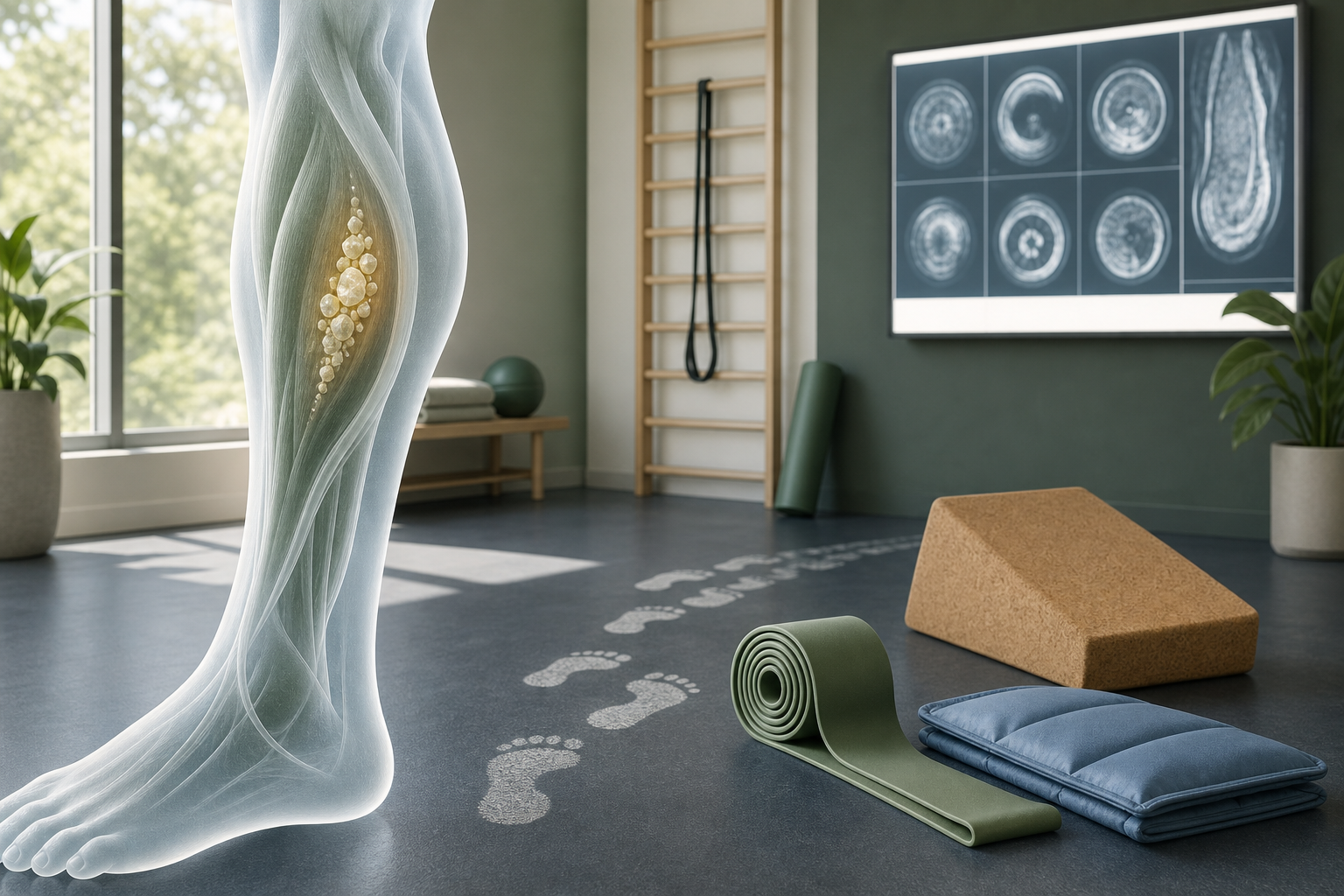
Treatment Options for Calcification of Muscle in the Left Lower Leg usually start with conservative care: activity modification, targeted physical therapy, soft tissue treatment, gait correction, and monitoring with imaging when needed. If you are asking what is muscle calcification in lower leg, the short answer is this: calcium can collect inside injured soft tissue, often after a significant strain, contusion, or repeated overload, and that can create pain, stiffness, and a firm area in the calf.
Most cases need a clear evaluation before you try to treat them on your own. A provider needs to distinguish muscle calcification from a calf strain, Achilles problem, nerve referral, or a vascular issue that needs urgent care.
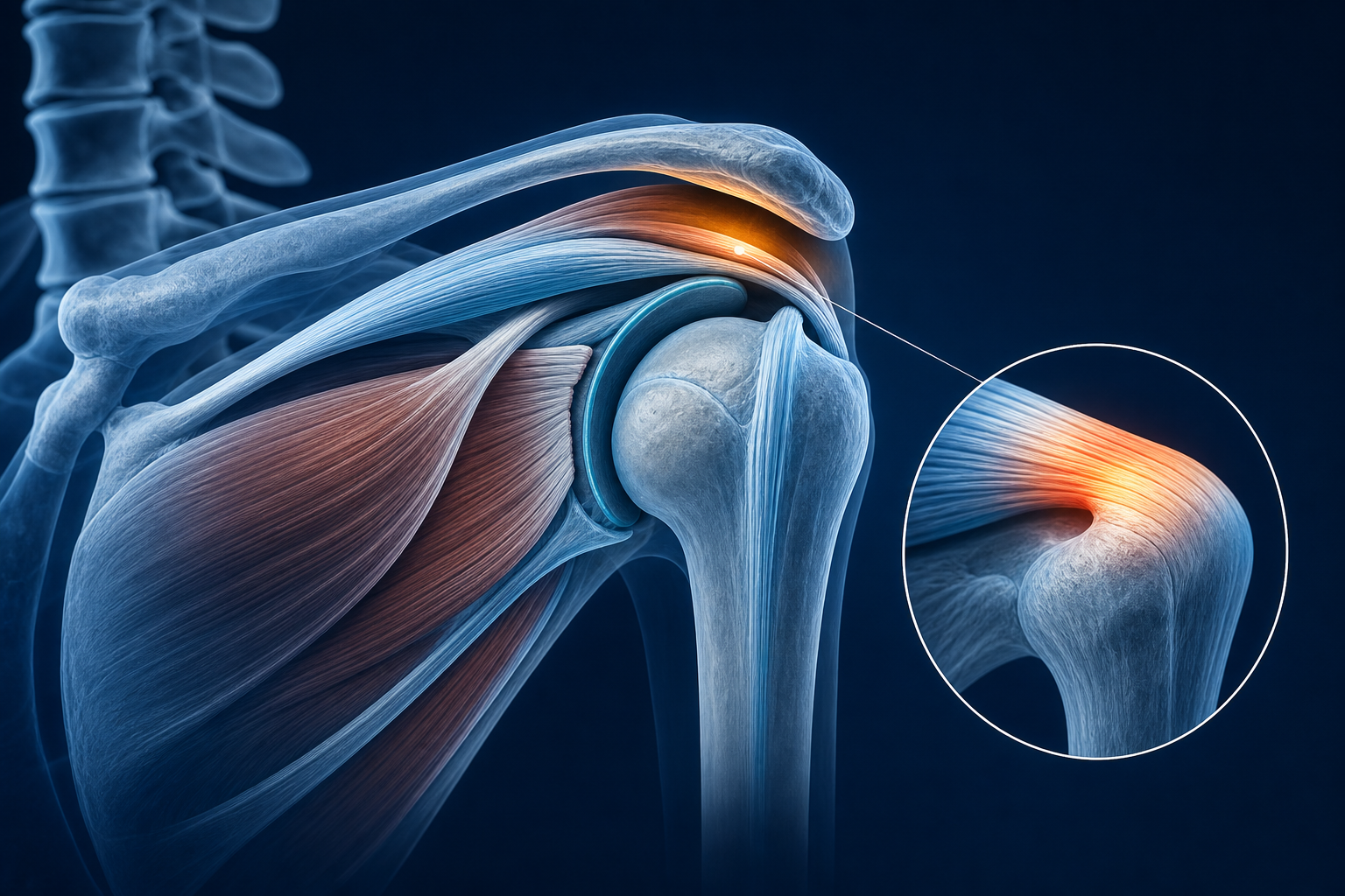
What Is Calcification of Muscle in the Left Lower Leg?
Muscle calcification means calcium deposits form within soft tissue that normally should stay flexible. In the left lower leg, this most often affects the calf region after trauma or repeated strain and may involve the gastrocnemius, soleus, or the connective tissue between muscle fibers.
This is not the same thing as simple muscle tightness.
When soft tissue heals abnormally, the body may lay down calcified material in or around injured fibers. That can limit ROM, change walking mechanics, and create a hard or gritty area that does not behave like a normal muscle knot. In some cases the process resembles myositis ossificans, where bone-like tissue forms inside muscle after a direct blow or deeper tissue injury.
- Normal muscle soreness: usually improves steadily over days
- Muscle strain: often painful with contraction and stretching, but texture remains mostly soft
- Calcification: may leave a persistent firm spot, restricted motion, and pain with loading weeks after the original injury
Research on post-traumatic soft tissue calcification suggests it is uncommon but well recognized after contusions and repetitive overload. If symptoms persist past the expected healing window, imaging is often considered.
Calf strains often improve in 2 to 6 weeks depending on grade. A firm mass, ongoing stiffness, or pain that is still limiting walking after that window deserves reassessment.
Where does calcification happen in the lower leg, and what does it affect?
In the lower leg, calcification usually shows up in the posterior compartment, especially the calf. The exact location matters because the medial gastrocnemius, lateral gastrocnemius, soleus, and Achilles tendon all load differently during walking, stairs, and push-off.
Common structures involved
- Gastrocnemius: the larger calf muscle that crosses the knee and ankle; often injured with sudden acceleration or jumping
- Soleus: deeper calf muscle that works heavily during walking and standing endurance
- Achilles region: not muscle, but nearby stiffness can change tendon loading
- Tibia and fibula borders: pain near these bones can confuse the picture and make providers consider other causes
- Tibial nerve pathway: nerve irritation can mimic calf symptoms but usually adds burning, tingling, or radiating patterns
Why left lower leg specificity matters
Left-sided symptoms are not treated differently just because they are on the left. What matters is whether your left ankle dorsiflexion is restricted, your foot strike is asymmetrical, or your pelvis and knee mechanics are shifting load into that calf. A provider may also compare the left and right side to measure swelling, tenderness, calf circumference, and ankle mobility.
Structure Typical symptom pattern What loading aggravates it Medial gastrocnemius Sharp pain with push-off, tenderness high in calf Running, lunging, fast walking Soleus Deep ache or tightness lower in calf Walking hills, prolonged standing, bent-knee loading Achilles region Stiffness near heel or lower calf Stairs, jumping, first steps after rest Calcified soft tissue area Firm lump, focal stiffness, persistent pain Stretching, compression, repeated calf contractionWhat symptoms suggest muscle calcification instead of routine calf tightness?
Pain and stiffness in left lower leg from calcification usually lasts longer than a routine strain and often includes a distinct firm area. If you notice a hard lump in calf after injury, reduced ankle motion, or pain that returns every time you increase activity, soft tissue calcification moves higher on the list.
The pattern usually looks like this:
- Initial injury, bruise, or strain
- Partial improvement over 1 to 3 weeks
- Persistent hard spot or deep stiffness that does not resolve
- Pain with calf raises, walking fast, stairs, or stretching
- Reduced dorsiflexion at the ankle
That said, not every hard spot is calcification. Providers also consider scar tissue, hematoma, fascial thickening, tendon involvement, and referred symptoms from the lumbar spine. If the pain travels from the buttock into the leg, compare that pattern with sciatica treatment and pain patterns, because nerve referral can mimic a calf problem.
A simple strain usually becomes more flexible as the weeks pass. Calcified tissue often stays mechanically stiff and locally tender even as swelling improves.
Seek urgent care immediately if you have sudden calf swelling, unusual warmth, skin color change, shortness of breath, or pain that is severe without a clear injury. Those signs need prompt medical assessment.
What causes calcification in left calf muscle after injury or repeated strain?
Calcification in left calf muscle causes usually fall into two buckets: a single significant injury or repeated overload that never fully settles. The common thread is tissue damage followed by disorganized healing.
A direct blow to the calf can create bleeding within the muscle. If the area is repeatedly compressed, stretched aggressively too early, or loaded before healing has matured, the repair process can become abnormal. Repeated sprinting, hill running, explosive sports, or altered gait from another problem can also keep stressing the same fibers.
Frequent contributing factors
- Contusion: a direct hit to the calf creates deeper tissue trauma.
- Moderate or severe strain: partial tearing of gastrocnemius or soleus fibers may heal with scar and calcified changes.
- Early return to sport: forceful stretching and loading too soon can irritate healing tissue.
- Poor ankle mobility: reduced dorsiflexion shifts more load into the calf during gait.
- Foot and knee mechanics: overpronation, limited great toe extension, or poor single-leg control can keep the calf overloaded.
The same biomechanical logic shows up in other regions of the body. Articles on non-pharmaceutical tension headache care and vertigo-related chiropractic support show a similar principle: restore mechanics first, then load tissue gradually.
How do providers diagnose muscle calcification?
If you are searching how do providers diagnose muscle calcification, the answer is straightforward: history, physical exam, movement testing, and imaging when the findings suggest more than a routine strain. Providers do not diagnose this from symptoms alone.
Your exam usually includes:
- Palpation for a firm or tender mass
- Measurement of ankle dorsiflexion and plantarflexion
- Calf strength testing, often with single-leg heel raises
- Walking and gait analysis
- Assessment of knee, hip, and foot mechanics
- Screening for nerve or vascular signs
X-ray may show mature calcification better than an early soft tissue lesion. Ultrasound can help visualize soft tissue texture and guide follow-up. MRI may be considered when the presentation is unclear or the provider needs to distinguish calcification from tear, hematoma, or another mass.
Imaging is more likely when symptoms last beyond 4 to 6 weeks, when a firm mass is growing, or when function is not improving.
Evaluation tool What it helps identify When it is commonly used Physical exam Tenderness, stiffness, weakness, gait changes First visit X-ray More mature calcified tissue Persistent symptoms or hard mass Ultrasound Soft tissue structure and healing changes Follow-up or unclear exam MRI Muscle injury detail and differential diagnosis Complex or non-improving casesTreatment Options for Calcification of Muscle in the Left Lower Leg
Treatment for muscle calcification in calf focuses on restoring motion, reducing mechanical irritation, and gradually reloading the tissue. Muscle calcification treatment without surgery often works best when the plan matches the stage of healing instead of forcing aggressive stretching into a stiff, irritated area.
Most conservative plans include several pieces at once.
- Reduce aggravating load: temporarily cut sprinting, jumping, hills, and long walks that flare symptoms.
- Restore ankle mobility: improve dorsiflexion so the calf is not overworking every step.
- Load the calf progressively: begin with pain-limited isometrics, then controlled raises, then single-leg work.
- Address scar and soft tissue restriction: manual therapy can help surrounding tissue glide better.
- Correct gait and kinetic chain issues: hip, knee, and foot mechanics matter.
- Monitor change over time: symptom reduction and improved ROM are better markers than chasing quick tissue changes.
Expected timelines vary. A mild persistent post-strain stiffness pattern may improve in 4 to 8 weeks of guided rehab. More established calcified lesions can take 2 to 6 months to become less limiting, especially if the area has been irritated for a long time.
Conservative care usually aims to improve function first: walking, stairs, calf strength, and ankle ROM. The tissue may remain palpable even after symptoms improve.
How can physical therapy and chiropractic care help?
Physical therapy for calf muscle calcification is usually the core of treatment because PT directly targets mobility, strength, and gait. Chiropractic care may support adjacent joint motion and biomechanics, especially if ankle, foot, knee, or pelvic mechanics are feeding the overload pattern.
Physical therapy priorities
- Restore ankle dorsiflexion
- Improve calf endurance with graded heel raise progressions
- Train eccentric control for walking downhill and deceleration
- Improve single-leg balance and foot control
- Correct compensations at the knee and hip
Can chiropractic help lower leg tightness?
Sometimes, yes. Chiropractic care does not remove calcification directly, but it may help when restricted joint motion is increasing calf load. A provider may assess the talocrural joint, subtalar joint, knee mechanics, and even lumbar referral patterns. If stiffness in the low back or nerve tension is contributing, that needs to be addressed alongside local calf rehab.
This same conservative model shows up across other conditions where mechanics matter, including upper cervical care discussions for Meniere’s-related dysfunction and natural rehabilitation strategies after concussion.
Approach Main goal Typical timeline Physical therapy ROM, strength, gait retraining 2-3 visits weekly for 3-6 weeks, then taper Chiropractic biomechanics support Joint motion and movement efficiency Varies by mobility findings Home exercise program Daily tissue loading and mobility gains Usually 6-12 weeks minimumSoft tissue care, stretching, and activity changes
Soft tissue treatment for calf calcification should be specific and measured. Deep, aggressive pressure directly into a highly irritable calcified area can flare symptoms. Better results usually come from treating the surrounding gastrocnemius, soleus, and fascial restrictions while rebuilding load tolerance.
Useful options may include:
- Manual soft tissue work around the restricted zone
- Instrument-assisted soft tissue techniques when tolerated
- Gentle mobility work for ankle dorsiflexion
- Heel raise progressions
- Temporary reduction in uphill walking, plyometrics, and sudden acceleration
Try this home routine once or twice daily if your provider says the tissue is safe to load:
- Walk for 3 to 5 minutes to warm the calf.
- Stand facing a wall with the affected leg behind you.
- Keep the back knee straight and heel down for a gastrocnemius stretch, 30 seconds x 3.
- Bend the back knee slightly for a soleus stretch, 30 seconds x 3.
- Perform double-leg calf raises, 2 sets of 10 slow reps.
- If pain stays mild, add 5 controlled lowering reps on the affected side.
- Finish with 5 ankle dorsiflexion rocks over the foot without lifting the heel.
Stop and get reassessed if the lump enlarges, pain sharply increases, or your walking worsens for more than 24 hours after exercise. For more condition education, explore more health topics.
What questions should you ask, and when should you see a provider?
If you are wondering is it normal to have calf pain after strain, the answer is yes for a short period, but not when pain stays focal, stiff, or mechanically limiting beyond the usual healing window. Ask better questions and you get a better treatment plan.
- Does this look like routine strain, scar tissue, or calcification?
- Do I need an X-ray, ultrasound, or MRI based on this exam?
- Which structure is involved: gastrocnemius, soleus, Achilles region, or something else?
- What movements should I avoid for the next 2 weeks?
- What benchmarks show I am improving: ROM, heel raises, walking speed, swelling, or pain?
- When should I be rechecked if the hard area does not change?
Routine appointment: lingering calf pain after 2 to 3 weeks, recurrent tightness, reduced ankle ROM, pain with stairs, or a persistent firm spot after injury.
Urgent assessment: sudden major swelling, redness, unusual heat, inability to bear weight, numbness, rapidly worsening pain, or shortness of breath.
What to Do Next
Start with a provider who treats lower-extremity biomechanics every day: a physical therapist, rehabilitation-focused chiropractor, or sports rehab provider. Your first visit should include a history of the injury, gait assessment, ankle and calf ROM testing, strength testing, and discussion of whether imaging is appropriate.
Bring specific details:
- When the pain started
- Whether there was a direct blow or strain
- Whether you noticed a hard lump in calf after injury
- What activities trigger pain: stairs, fast walking, running, or stretching
- How long symptoms have lasted
If you need a rehabilitation provider for lower leg pain near me, use Medximity to find a physical therapist near you, find a chiropractor near you, or browse providers. The right plan usually combines diagnosis, guided loading, and movement correction rather than repeated rest alone.
Persistent left lower leg stiffness has a mechanical reason. Get it measured, identify the tissue involved, and follow a step-by-step rehab plan that restores walking, calf strength, and ankle motion.