Living with Sprain of tibiofibular ligament of right ankle, initial encounter: A Comprehensive Guide means you’re dealing with a “high ankle sprain” (a syndesmosis sprain) that typically heals slower than a common lateral ankle sprain. Your priorities in the first 7–14 days are protecting the distal tibiofibular syndesmosis, controlling swelling, and avoiding early twisting that separates the tibia and fibula. Most mild-to-moderate syndesmosis sprains improve with structured rehab over 6–12 weeks, with return to running/cutting often closer to 8–12+ weeks depending on severity and exam findings.
Understanding Your Tibiofibular Ligament Sprain
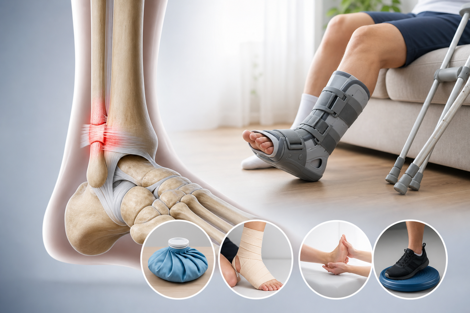
A tibiofibular ligament sprain involves the syndesmotic ligaments that hold the bottom of your tibia and fibula together, forming the “mortise” that the talus sits in. The main structures are the anterior inferior tibiofibular ligament (AITFL), posterior inferior tibiofibular ligament (PITFL), and the interosseous ligament/membrane. When these tissues are strained, the ankle can feel unstable with rotation, and pushing off can be sharply limited.
High ankle sprains usually happen with the foot planted and the leg rotating inward (external rotation of the foot relative to the tibia), or with forced dorsiflexion. This mechanism stresses the AITFL first, then deeper syndesmotic tissues if the force continues.
- Typical location of pain: front/outside of the ankle above the ankle joint line (over the AITFL), sometimes spreading up the lower leg along the interosseous membrane.
- Movements that flare symptoms: pivoting, cutting, deep squat, stepping off a curb, walking fast, climbing stairs.
- Why it matters: the syndesmosis is a “stability ring.” If it gapes under load, you can’t push off efficiently, and rehab must emphasize protection early.
High ankle sprains generally take longer to return to sport than lateral ankle sprains, often requiring multiple weeks of progressive rehabilitation. Source: American Academy of Orthopaedic Surgeons (AAOS), “High Ankle Sprain (Syndesmosis Sprain)”.
Is This a High Ankle Sprain (Syndesmosis) or a Common Lateral Ankle Sprain?
A syndesmosis sprain hurts higher and is provoked by rotation; a lateral ankle sprain is usually more below the ankle bone and is provoked by inversion (rolling inward). You can’t self-diagnose severity reliably, but you can use pattern recognition to decide how urgently to be evaluated.
Quick pattern check you can do safely
- Syndesmosis pattern: pain above the ankle joint line; pain with turning/pivoting; discomfort with “knee over toes” dorsiflexion; pain with squeezing the lower leg can occur.
- Lateral sprain pattern: pain and swelling around the outside ankle bone (lateral malleolus), especially over the anterior talofibular ligament (ATFL); bruising around the ankle/foot; pain mostly with rolling inward.
Red flags: get urgent evaluation today
Use these as “do not wait” criteria. They are not rare with ankle injuries.
- Inability to take 4 steps right after injury or in the clinic setting.
- Bone tenderness at the back edge/tip of the lateral malleolus or medial malleolus, or at the base of the 5th metatarsal.
- Visible deformity, rapidly expanding swelling, or numb/cold foot.
- Severe pain above the ankle with rotation plus a feeling the ankle is “spreading.”
The Ottawa Ankle Rules help determine when imaging is indicated after ankle injury and are widely used to reduce missed fractures. Source: American College of Radiology (ACR) Appropriateness Criteria: Acute Trauma to the Ankle.
Immediate Conservative Care (First 72 Hours): What Actually Helps
Protecting the syndesmosis early prevents repeated micro-separation of the tibia and fibula while the AITFL and interosseous tissues start to repair. Your default should be relative rest + protection + swelling control, not aggressive stretching.
Protection and load management
- Brace/boot: Use a supportive ankle brace or walking boot if walking increases pain above a 3/10. A boot limits rotation better than a soft brace.
- Crutches: Use them if you limp. Limping repeats torsion at the syndesmosis.
- Activity rule: If an activity increases pain during the activity and stays elevated the next morning, it was too much.
Swelling control you can measure
- Compression: Use an elastic wrap from mid-foot to mid-calf, snug but not numb/tingly.
- Elevation: 20–30 minutes, 3–5 times/day, ankle above heart level.
- Movement “pumps”: 30–60 gentle ankle pumps every 1–2 hours while awake (pain-free range).
Skip early aggressive calf stretching if it creates a sharp pinch above the ankle. That often means you’re driving dorsiflexion and rotation into irritated syndesmotic fibers.
Early protected motion and progressive loading are commonly used principles in ankle ligament rehabilitation to restore function while controlling swelling. Source: Journal of Orthopaedic & Sports Physical Therapy (JOSPT) Clinical Practice Guidelines for Ankle Stability and Movement Coordination Impairments.
What Rehab Looks Like Week-by-Week for a Right High Ankle Sprain
Most conservative plans follow phases: (1) protect and calm symptoms, (2) restore ROM, (3) rebuild strength and proprioception, (4) return to impact and cutting. The right ankle detail matters because driving and stepping patterns often load the right side more during braking and pivoting.
- Days 1–7: prioritize protection. Goal: walk short distances without a limp (with brace/boot if needed). ROM: ankle pumps and gentle circles in pain-free range.
- Weeks 2–4: ROM and controlled strength. Goal: normal walking mechanics, improved dorsiflexion without pinch above the ankle. Begin balance drills when weight-bearing is comfortable.
- Weeks 4–8: strength + neuromuscular control. Goal: single-leg balance 30–45 seconds, controlled calf raises, step-down control without collapse.
- Weeks 8–12+: graded return to running/jumping/cutting. Goal: hop and change-of-direction drills with minimal next-day symptoms.
Two timelines you can use to sanity-check progress:
- Walking timeline: many mild-to-moderate cases regain near-normal walking in 2–4 weeks with protection and structured rehab.
- Sport timeline: running and pivoting often take 8–12+ weeks because syndesmotic tissues dislike rotation under load.
High ankle sprains often require longer rehabilitation than lateral ankle sprains, with return-to-sport commonly extending beyond several weeks depending on severity. Source: AAOS OrthoInfo (High Ankle Sprain).
Home Exercise Protocol (Step-by-Step) for the First 2–4 Weeks
Do these drills to restore ankle motion and control while limiting the rotation that irritates the syndesmosis. Stop any drill that causes a sharp pain above the ankle joint line or causes swelling to spike later that day.
Phase 1 (Days 1–10): restore motion without torsion
- Ankle pumps: sitting or lying down, move foot up/down 30–60 reps, 4–6 times/day.
- Alphabet (small letters): draw A–Z with your toes using small motions, 1–2 rounds/day.
- Isometric eversion/inversion: press the outside of your foot gently into a wall (eversion) for 10 seconds x 5 reps; repeat pressing inside of foot (inversion). Keep ankle neutral; don’t twist.
Phase 2 (Weeks 2–4): strength + balance (if walking is improving)
- Double-leg calf raises: hold a counter, rise up 2 seconds, lower 3 seconds, 2 sets of 10–15. Progress to more load only if no next-day flare.
- Theraband dorsiflexion: pull foot up against band, 2–3 sets of 12–15. Keep knee bent slightly to reduce joint pinch.
- Supported single-leg balance: stand on right foot near a counter, fingertip support allowed, 3 rounds of 20–45 seconds. Keep knee over 2nd toe; don’t let arch collapse.
One simple progression rule: increase total reps or time by 10–20% per week if next-day symptoms are stable.
Exercise therapy and balance training reduce recurrent ankle problems and improve function after ankle sprain. Source: JOSPT Clinical Practice Guidelines (Ankle Stability and Movement Coordination Impairments).
Which Conservative Treatments Work Best, and How Long Do They Take?
The best plan combines protection (brace/boot), progressive rehab, and manual therapy when appropriate. Passive treatments alone rarely restore the strength and proprioception you need for uneven ground and quick turns.
Treatment Best for What you should notice Typical timeline Walking boot (short-term) Moderate pain with walking; pain above ankle with rotation Less pain during walking; reduced swelling by end of day Often 1–3 weeks, then transition to brace as tolerated Lace-up brace or semi-rigid brace Transition back to normal shoes; limiting rotation More stable walking; fewer “tweaks” on turns Commonly 3–8+ weeks depending on activity Physical therapy (strength, balance, return-to-run) Restoring ROM, calf strength, peroneal control, landing mechanics Improved dorsiflexion, better single-leg control, reduced next-day soreness Often 6–12 sessions over 4–8 weeks; longer for athletes Chiropractic care (ankle/foot mechanics, joint mobility) Stiff talocrural/subtalar joints; gait compensation up the chain Smoother gait; improved ankle dorsiflexion tolerance Commonly 2–6 weeks alongside rehab Manual therapy + soft tissue work Calf tightness (gastrocnemius/soleus), peroneal guarding, swelling management Less stiffness; improved ROM for squatting/steps Often starts week 2+, progresses with tolerance Return-to-sport progression (graded running, hops, cutting) Field/court sports; jobs requiring pivots/ladders No sharp syndesmosis pain; minimal swelling next day Frequently 8–12+ weeks- ROM target that matters: compare knee-to-wall dorsiflexion right vs. left. A difference >2–3 cm often correlates with ongoing functional limits.
- Strength target that matters: controlled single-leg calf raises. Many people need to rebuild to 20–25 reps before higher-level impact feels normal.
How Do You Modify Walking, Work, Driving, and Exercise Without Slowing Recovery?
You heal faster when you keep the ankle loaded within tolerance and avoid repeated torsion. “No movement” often leads to stiffness; “too much twisting” keeps the syndesmosis irritated.
- Walking: shorten your stride and keep toes pointed forward. Avoid fast turns; use step-turns (move feet, don’t pivot on the injured foot).
- Stairs: go up leading with the uninjured leg; go down leading with the injured leg while holding the rail. This reduces dorsiflexion torque on the right ankle.
- Work: if you stand for long periods, schedule 5-minute “elevation breaks” every 60–90 minutes for the first 1–2 weeks.
- Driving (right ankle): avoid driving if braking causes sharp pain or delayed swelling. Practice gentle ankle pumps before driving to reduce stiffness; stop if reaction time feels limited.
- Cardio options: upper-body ergometer, seated strength circuits, or cycling only if it’s pain-free and doesn’t increase next-day swelling. Avoid rowing early because it forces repeated dorsiflexion.
Persistent swelling, loss of motion, and impaired balance are common after ankle sprain and are key targets for rehabilitation to reduce recurrence. Source: National Library of Medicine / PubMed overview of ankle sprain rehabilitation concepts and recurrence risk.
Use a simple daily self-check:
- Morning pain score (0–10)
- Swelling compared to yesterday (same/better/worse)
- Walking quality (normal/limp)
If two of the three worsen for 48 hours, reduce loading and get reassessed.
What to Do Next
Get evaluated if you have pain above the ankle joint line, pain with rotation, or you can’t walk without limping after 3–5 days. A provider should assess the syndesmosis (AITFL/PITFL), check the talocrural and subtalar joints, screen the peroneal tendons, and confirm whether imaging is needed using established rules (ACR/Ottawa).
- Best-fit providers: a physical therapist for staged rehab and return-to-run testing; a chiropractor for ankle/foot mechanics and mobility work when appropriate; sports rehab providers for balance and agility progression.
- What a first visit should include: gait assessment, swelling measurement, dorsiflexion testing (knee-to-wall), ligament stress tests as tolerated, balance testing, and a written home program with progression criteria.
- Seek urgent care now if you cannot take 4 steps, have deformity, numb/cold foot, rapidly increasing swelling, or bone tenderness at the malleoli or base of the 5th metatarsal.
- Routine care (next available appointment) if you can walk but you have persistent pain with turning, limited dorsiflexion, or repeated “tweaks” on uneven ground.
Use Medximity tools to decide and act:
- check your symptoms for next-step guidance based on what you can and can’t do today.
- find a physical therapy near you for structured syndesmosis rehab and return-to-activity testing.
- find a chiropractor near you for mobility, gait mechanics, and conservative ankle care.
- browse providers if you want to compare specialties and availability.
- explore more health topics for related guides on ankle stability, balance training, and safe return to activity.
FAQ
How long does a tibiofibular ligament (high ankle) sprain take to heal?
Mild-to-moderate cases often improve enough for near-normal walking in 2–4 weeks, but return to running, jumping, and cutting commonly takes 8–12+ weeks. Your timeline depends on pain with rotation, exam stability, swelling response, and how quickly dorsiflexion and calf strength return.
Should you keep walking on a high ankle sprain?
Walk only if you can do it without a limp or with support (brace/boot) that keeps pain ≤3/10. Limping and pivoting repeatedly stress the syndesmosis. Use crutches temporarily if needed and progress weight-bearing as next-day swelling and pain allow.
What exercises should you avoid early on?
Avoid drills that combine deep dorsiflexion with rotation, including aggressive calf stretching that causes a pinch above the ankle, pivoting, cutting, and uneven-ground hikes. Start with ankle pumps, gentle ROM, and isometrics, then add calf raises and balance once walking mechanics normalize.
When do you need imaging for an ankle sprain?
Imaging is commonly recommended when you cannot take 4 steps, have bone tenderness at key points (malleoli, base of 5th metatarsal), deformity, or concerning neurovascular signs. Clinicians often apply the Ottawa Ankle Rules and ACR guidance to decide. Sources: ACR Appropriateness Criteria (Acute Trauma to the Ankle) and AAOS OrthoInfo.
Can physical therapy help a high ankle sprain?
Yes. PT targets dorsiflexion ROM, calf and peroneal strength, balance/proprioception, and return-to-run criteria. A common plan is 6–12 visits over 4–8 weeks, adjusted based on swelling and functional testing.
What are signs your high ankle sprain is not improving normally?
Red flags for reassessment include persistent inability to walk without limping after 10–14 days, worsening swelling for 48 hours despite load reduction, sharp pain above the ankle with light pivoting, or repeated giving-way episodes. These patterns warrant a clinician exam to reassess syndesmosis stability and rule out associated injuries.