Understanding Lumbosacral Spinal Stenosis: What You Need to Know
Lumbosacral spinal stenosis is a condition where the spinal canal narrows in the lower back region, potentially causing compression of nerve structures. This narrowing occurs specifically in the lumbosacral area, which includes the five lumbar vertebrae and the sacrum at the base of the spine. Understanding this condition becomes crucial as it affects millions of adults, particularly those over 50, and can significantly impact quality of life and mobility.
The importance of learning about lumbosacral spinal stenosis extends beyond just medical knowledge—it empowers patients to recognize symptoms early, understand treatment options, and make informed decisions about their care. This comprehensive guide will walk through seven essential facts about the condition, covering everything from anatomy and symptoms to treatment approaches and long-term management strategies, helping patients navigate their healthcare journey with confidence.
1. What Lumbosacral Spinal Stenosis Actually Means for Your Body
The lumbosacral spine represents the junction between the lower back and pelvis, consisting of the five lumbar vertebrae (L1-L5) and the sacrum. This region bears significant weight and stress during daily activities, making it particularly vulnerable to degenerative changes over time. The spinal canal in this area houses the cauda equina, a bundle of nerve roots that control sensation and function in the legs, bladder, and bowel.
When stenosis develops, the space within the spinal canal becomes narrowed due to various factors such as bone spurs, thickened ligaments, or bulging discs. This narrowing can compress the nerve structures, leading to the characteristic symptoms patients experience. Unlike a herniated disc that typically affects a single nerve root, lumbosacral spinal stenosis often affects multiple nerve pathways simultaneously.
The condition differs significantly from other spinal problems because it typically develops gradually and symptoms may worsen with certain positions or activities. While conditions like acute disc herniation often cause sudden, sharp pain, spinal stenosis symptoms tend to develop slowly and may be relieved by sitting or leaning forward, which opens up the spinal canal slightly.
2. Recognizing the Warning Signs and Symptoms
Neurogenic claudication represents the hallmark symptom of lumbosacral spinal stenosis, characterized by pain, weakness, or cramping in the legs that occurs with walking or standing. Patients often experience these symptoms after walking a certain distance, which gradually decreases as the condition progresses. The discomfort typically improves when sitting down or leaning forward, such as when pushing a shopping cart.
Pain patterns in lumbosacral spinal stenosis can vary significantly between individuals. Some patients experience aching or burning sensations that radiate from the lower back into the buttocks and legs. Others may notice numbness, tingling, or a feeling of heaviness in their legs. The symptoms are often bilateral, affecting both legs, though they may be more pronounced on one side.
The impact on daily activities becomes increasingly noticeable as the condition progresses. Patients may find themselves unable to walk the distances they once could, leading to decreased activity levels and potential deconditioning. Simple tasks like grocery shopping, walking the dog, or climbing stairs may become challenging. Many patients develop compensatory behaviors, such as using shopping carts for support or taking frequent breaks during activities that require prolonged standing or walking.
3. Understanding the Root Causes and Risk Factors
Age-related degenerative changes represent the primary cause of lumbosacral spinal stenosis, with the natural aging process leading to wear and tear on spinal structures. As people age, the ligaments in the spine may thicken, bone spurs can develop on the vertebrae, and the discs between vertebrae may lose height and bulge outward. These changes collectively contribute to narrowing of the spinal canal over time.
Several contributing conditions can accelerate or worsen spinal stenosis development. Spondylolisthesis, where one vertebra slips forward over another, can significantly reduce canal space. Degenerative disc disease, arthritis of the facet joints, and previous spinal injuries or surgeries may also contribute to stenosis development. Additionally, some individuals may be born with a naturally narrow spinal canal, making them more susceptible to symptoms as degenerative changes occur.
Lifestyle and genetic factors play important roles in stenosis development and progression. Occupations requiring heavy lifting, repetitive bending, or prolonged sitting may increase risk over time. Family history of spinal problems can indicate genetic predisposition to degenerative changes. Other factors such as smoking, obesity, and sedentary lifestyle may accelerate the degenerative process, while maintaining good posture, regular exercise, and healthy body weight may help slow progression.
4. How Healthcare Providers Assess Lumbosacral Spinal Stenosis
Physical examination techniques employed by healthcare providers focus on identifying characteristic signs of neurogenic claudication and nerve compression. Providers may assess walking tolerance, observe posture changes, and perform specific tests such as having patients walk until symptoms appear, then sit to see if symptoms improve. Neurological testing includes checking reflexes, muscle strength, and sensation in the legs to identify areas of nerve involvement.
Imaging tests serve crucial roles in confirming the presence and extent of spinal stenosis. Magnetic resonance imaging (MRI) provides detailed views of soft tissues, including nerves, discs, and ligaments, allowing providers to see the degree of canal narrowing and nerve compression. CT scans may be used to better visualize bone structures and spurs. X-rays can show alignment issues, bone spurs, and signs of degenerative changes, though they cannot directly show nerve compression.
Important questions patients should consider asking during evaluation include: "What is the severity of my stenosis?" "Which nerve structures are affected?" "What treatment options might be most appropriate for my situation?" "How might this condition progress over time?" "What lifestyle modifications might help manage symptoms?" Understanding these aspects helps patients participate more actively in their care planning and make informed decisions about treatment approaches.
5. Non-Operative Treatment Options That May Help
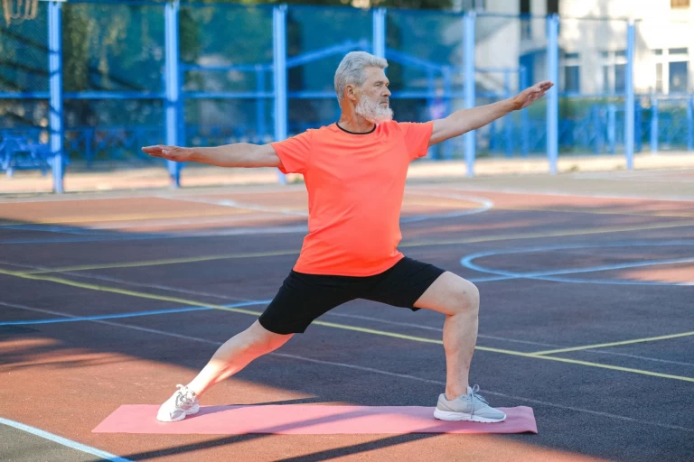
Physical therapy approaches form the cornerstone of conservative management for lumbosacral spinal stenosis. Therapists may focus on exercises that promote spinal flexion, as this position can help open the spinal canal and reduce nerve compression. Core strengthening exercises can improve spinal stability, while cardiovascular conditioning helps maintain overall fitness despite walking limitations. Aquatic therapy may be particularly beneficial as the buoyancy of water reduces stress on the spine.
Medication management options that healthcare providers may consider include anti-inflammatory medications to reduce swelling around compressed nerves, though their effectiveness for stenosis symptoms varies. Some patients may benefit from medications that address nerve-related pain. Topical treatments or muscle relaxants might also be considered depending on individual symptoms and overall health status.
Epidural steroid injections and other interventions represent additional options for symptom management. These procedures involve injecting anti-inflammatory medication directly into the epidural space around compressed nerves, potentially providing temporary symptom relief. Other interventions might include nerve blocks or trigger point injections. The effectiveness of these treatments varies among individuals, and healthcare providers typically consider them as part of a comprehensive treatment approach rather than standalone solutions.
6. When Operative Intervention Becomes Necessary and What to Expect
Indications for operative intervention typically arise when conservative treatments have not provided adequate symptom relief and the condition significantly impacts quality of life. Healthcare providers may consider operative options when patients experience progressive weakness, severe walking limitations, or bladder/bowel dysfunction. The decision involves careful consideration of the patient's overall health, symptom severity, and personal goals for treatment outcomes.
Types of decompression procedures commonly performed include laminectomy, where portions of the vertebral bone are removed to create more space for nerves, and laminotomy, a less extensive procedure targeting specific areas of compression. Some patients may require fusion procedures if spinal instability is present. Minimally invasive techniques have evolved to reduce tissue disruption while achieving decompression goals, potentially leading to faster recovery times.
Recovery timeline and expectations vary significantly based on the specific procedure performed and individual patient factors. Initial recovery typically involves a hospital stay of one to several days, followed by a gradual return to activities over several weeks to months. Physical therapy often plays an important role in post-operative recovery. While many patients experience significant improvement in leg symptoms, complete symptom resolution may not always occur, and realistic expectations should be established before any procedure.
7. Living with Stenosis: Lifestyle Changes and Long-term Management
Exercise modifications and recommendations focus on activities that allow forward flexion of the spine, which can help alleviate symptoms. Stationary bicycling, walking with support (such as using a walker), and swimming often work well for maintaining cardiovascular fitness. Activities that require prolonged standing or walking without support may need to be modified or avoided during symptom flares.
Ergonomic considerations can significantly impact daily comfort and function. Using shopping carts for support while walking, adjusting work stations to promote good posture, and choosing supportive seating can help manage symptoms throughout the day. Some patients find that adjustable beds or reclining chairs provide better comfort for sleeping and resting.
Prevention strategies for progression include maintaining a healthy weight to reduce stress on the spine, staying as active as possible within symptom limitations, and avoiding activities that consistently worsen symptoms. Regular follow-up with healthcare providers helps monitor the condition's progression and adjust treatment approaches as needed. Staying informed about the condition and maintaining open communication with the healthcare team supports optimal long-term management and quality of life.
Taking the Next Step in Your Care Journey
Understanding lumbosacral spinal stenosis empowers patients to recognize symptoms, explore treatment options, and make informed healthcare decisions. The condition affects each person differently, making personalized care essential for optimal outcomes. From conservative management approaches to potential operative interventions, multiple options exist to help patients maintain their quality of life and functional abilities.
Professional medical care remains crucial for proper evaluation and treatment planning. Healthcare providers can assess individual situations, recommend appropriate treatments, and monitor progression over time. Finding qualified specialists who understand spinal conditions can make a significant difference in treatment outcomes and patient satisfaction. Taking an active role in your healthcare journey, asking questions, and staying informed about your condition supports the best possible outcomes for managing lumbosacral spinal stenosis.
This information is for educational purposes only and should not replace professional medical advice. Always consult with a qualified healthcare provider for personalized medical guidance.