Knee Subluxation: What Patients Need to Know: a knee subluxation is a partial shift of the knee joint, most often the patella moving partly out of its groove on the femur and then sliding back in. It differs from a full dislocation because the joint surfaces still keep some contact. Conservative care usually focuses on swelling control, restoring ROM, strengthening the quadriceps and hip muscles, and correcting movement patterns that let the knee give way.
What Is Knee Subluxation?
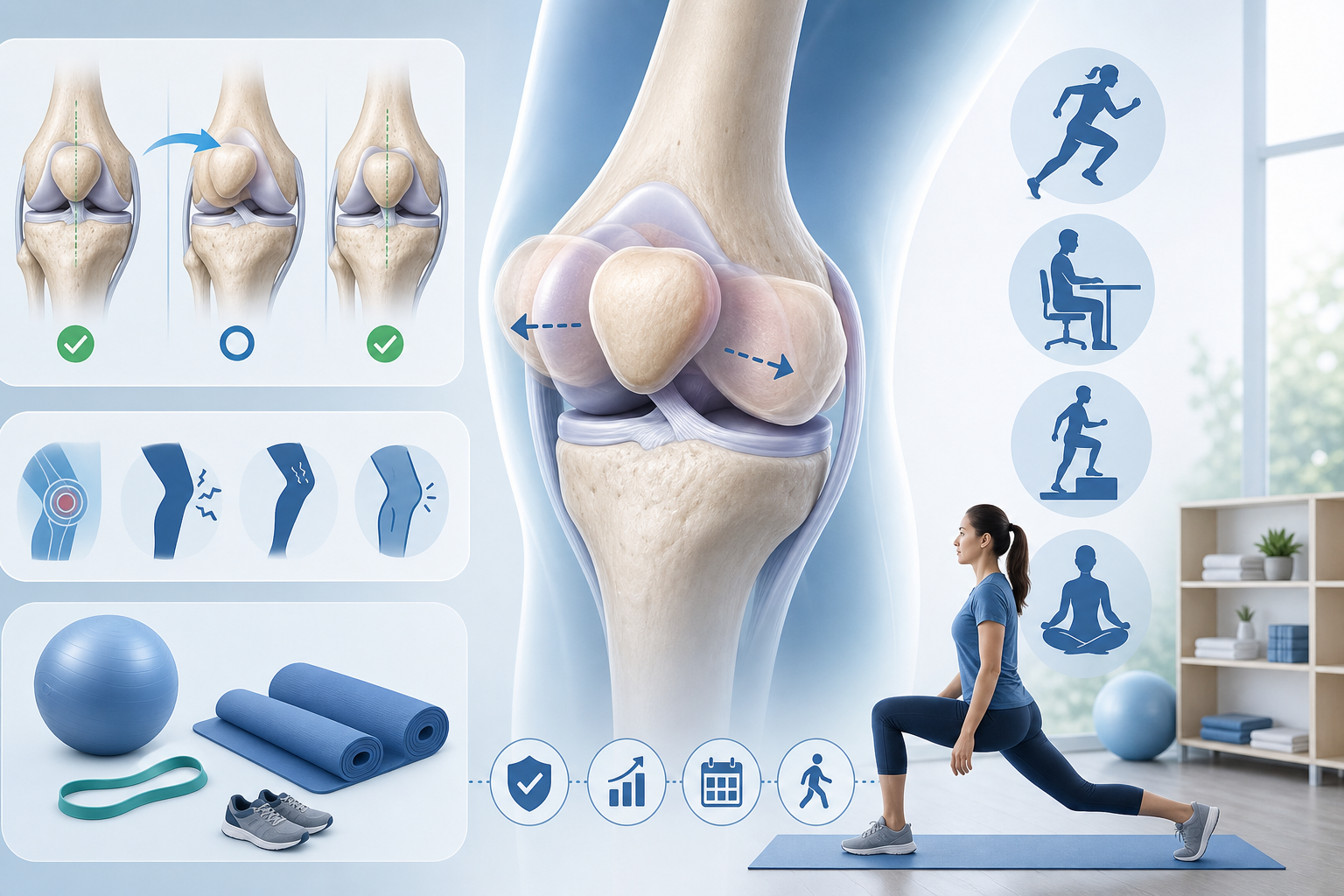
Knee subluxation means part of the knee joint briefly loses normal alignment without a complete joint separation. The most common version is patellar subluxation, often described as the kneecap slipping out of place without full dislocation. The patella usually shifts toward the outside of the knee, away from the midline, then returns to its track in the trochlear groove of the femur.
A less common but more serious pattern is tibiofemoral subluxation, where the tibia shifts partly against the femur. This pattern can stress the ACL, PCL, collateral ligaments, meniscus, and nearby blood vessels or nerves. A full tibiofemoral dislocation needs urgent evaluation because circulation below the knee can be affected.
Patellar instability research commonly reports recurrence after a first lateral patellar instability event in roughly 15% to 40% of cases, with higher risk in younger athletes and people with joint laxity.
If you are searching “what is a knee subluxation and how is it treated,” the short answer is: identify which joint surface is shifting, rule out serious injury, reduce swelling, restore motion, then build strength and control around the knee, hip, ankle, and foot.
- Partial shift: joint surfaces move out of normal alignment but do not fully separate.
- Full dislocation: joint surfaces fully lose contact and may stay displaced.
- Instability: the knee feels like it may buckle, slide, or give way during movement.
For a broader explanation of how subluxation is discussed in chiropractic care, see what an upper cervical subluxation means.
Patellar Subluxation vs. Tibiofemoral Subluxation: What Is the Difference?
The key difference between patellar and tibiofemoral subluxation is the structure that shifts. In patellar subluxation, the kneecap partially slips out of the femoral groove. In tibiofemoral subluxation, the main hinge joint between the tibia and femur partially shifts.
Patellar subluxation is more common in sports, stair climbing, squatting, and quick direction changes. It often involves the medial patellofemoral ligament, or MPFL, which helps prevent the patella from sliding too far outward. The vastus medialis oblique, or VMO, also helps guide the kneecap during knee extension.
Tibiofemoral subluxation usually involves higher force or significant ligament instability. The ACL, PCL, medial collateral ligament, lateral collateral ligament, meniscus, and posterolateral corner may need assessment.
Type What Moves Common Description Typical Concern Patellar subluxation Patella shifts on the femur Kneecap slips out and back in MPFL irritation, quad weakness, tracking problems Tibiofemoral subluxation Tibia shifts against the femur Knee shifts or buckles deeply Major ligament, meniscus, nerve, or blood vessel stress Full dislocation Joint surfaces fully separate Knee or kneecap stays out of place Urgent evaluation needed, especially with numbness or poor circulationFor the question “knee subluxation vs full dislocation what is the difference,” remember this rule: subluxation partially shifts, dislocation fully separates. Severe trauma, visible deformity, foot numbness, pale or cold skin, or inability to bear weight should be treated as urgent.
What Causes Knee Subluxation in Athletes and Non-Athletes?
Knee subluxation usually comes from a mix of anatomy, tissue laxity, strength deficits, and movement mechanics. Young athletes often sublux during cutting, pivoting, landing from a jump, or contact to the side of the knee. Sedentary adults may notice instability when standing from a chair, walking downhill, stepping off a curb, or twisting while the foot is planted.
Traumatic knee subluxation
Traumatic subluxation happens after a clear event. Examples include a fall, sports collision, awkward landing, or sudden twist. Swelling within 2 to 6 hours after injury suggests deeper joint irritation and should be evaluated, especially if the knee will not fully straighten.
Chronic or recurrent knee subluxation
Chronic knee subluxation means the knee repeatedly slips, shifts, or gives way. Recurrent episodes often relate to hip weakness, poor patellar tracking, ligament laxity, flat feet, ankle stiffness, or prior trauma that never fully regained strength and control.
- Ligament laxity: looser connective tissue allows extra joint motion.
- Muscle imbalance: weak VMO, gluteus medius, or hamstrings can alter knee tracking.
- Hypermobility: increased joint range may reduce passive stability.
- Prior injury: earlier sprains or patellar instability can increase recurrence risk.
- Foot and ankle mechanics: overpronation or limited ankle dorsiflexion can push the knee inward.
Patients with foot mechanics that affect knee tracking may also benefit from reading about chiropractic care for plantar fasciitis, since the arch, ankle, tibia, and knee work as one chain during walking.
Symptoms of Knee Subluxation: How Can You Tell?
Knee subluxation often feels like a quick slip, shift, pop, or buckle followed by pain around the kneecap or deeper in the joint. The phrase “knee gives way when walking causes and treatment” usually points to instability that needs a movement exam, not just rest.
If you are asking “is my kneecap partially dislocated how to tell,” look for a pattern: the patella moves outward during a twist, squat, stair descent, or athletic cut, then returns to place. You may notice swelling, tenderness along the inner edge of the kneecap, reduced confidence on stairs, or pain when sitting with the knee bent.
Patient checklist before your visit
- Did the knee shift outward, inward, backward, or feel like it rotated?
- Did swelling appear within the first 2 to 6 hours?
- Can you fully straighten and bend the knee?
- Can you walk 4 steps without the knee buckling?
- Do you feel pain along the kneecap, joint line, calf, or back of the knee?
- Did you hear or feel a pop during the injury?
- Has this happened before?
Seek urgent care now if the knee looks deformed, the foot becomes cold or pale, you develop numbness or weakness below the knee, calf swelling increases quickly, or you cannot bear weight after trauma.
Head or neck symptoms after a fall need separate attention. Review what to do after a possible concussion and why neck injuries should be taken seriously if the knee injury happened during a collision or fall.
Conservative Treatment Options for Knee Subluxation
Conservative treatment for knee subluxation targets swelling, ROM, patellar tracking, hip control, and return-to-activity strength. Chronic knee subluxation treatment without operative care usually works best when the plan addresses the cause: weak hip abductors, poor landing mechanics, tight lateral tissues, ankle stiffness, or general hypermobility.
Common treatment tools
- Activity modification: reduce pivoting, deep squats, running hills, and jumping for 1 to 3 weeks if symptoms flare.
- Compression and elevation: control swelling so the quadriceps can contract normally.
- Manual therapy: improve patellar mobility, ankle dorsiflexion, hip mobility, and soft tissue tone in the quadriceps, IT band, calves, and hamstrings.
- Exercise therapy: strengthen the VMO, gluteus medius, gluteus maximus, hamstrings, and calf complex.
- Balance training: retrain single-leg control on stable surfaces before uneven surfaces.
- Bracing or taping: support patellar tracking during early activity when appropriate.
Home exercise: quad set to straight-leg raise progression
- Lie on your back with the injured knee straight and the other knee bent.
- Tighten the front thigh muscle by pressing the back of the knee gently toward the floor.
- Hold 5 seconds, then relax 5 seconds. Repeat 10 times.
- If you can tighten the quad without pain or lag, lift the straight leg 12 inches.
- Lower slowly over 3 seconds. Perform 2 sets of 8 to 12 reps.
- Stop if the kneecap feels like it is sliding, pain sharpens, or swelling increases later that day.
To start care with the right provider, you can find a physical therapist near you or find a chiropractor near you.
What Happens During a Chiropractic or Rehabilitation Evaluation?
A knee subluxation evaluation checks whether the problem is isolated to the patella or involves deeper knee instability. A chiropractor or rehabilitation provider will usually start with mechanism of injury, swelling timing, prior instability, sports or work demands, and whether the knee locks, catches, or gives way.
The exam often compares both knees. Expect ROM testing, gait observation, squat or step-down assessment, patellar glide testing, ligament stress tests, and hip/ankle mobility checks. The provider may assess the gluteus medius, VMO, hamstrings, calves, foot arch control, and balance on one leg. If red flags appear, the provider may refer you for imaging or urgent medical evaluation.
What your provider may test
- Patellar apprehension: checks whether the kneecap feels unstable when guided outward.
- Single-leg squat: shows hip control, knee valgus, and arch collapse.
- Step-down test: reveals pain or poor tracking during stair-like loading.
- Lachman and drawer testing: screens ACL and PCL stability.
- Joint line palpation: checks meniscus-related tenderness.
If you search “chiropractor for knee subluxation near me,” choose a provider who evaluates the hip, ankle, foot, and spine along with the knee. The knee rarely fails in isolation. Pelvic control, ankle dorsiflexion, and foot mechanics can change how the tibia rotates under the femur.
For lower-body nerve symptoms that travel from the back into the leg, see what can be done for sciatic pain, since nerve irritation can mimic weakness or leg instability.
Frequently Asked Questions About Knee Subluxation
Most knee subluxation questions come down to safety, recurrence risk, and timeline. These quick answers help you decide whether to rest, schedule conservative care, or seek urgent evaluation.
How long does knee subluxation take to heal?
Mild patellar subluxation may calm down in 1 to 3 weeks, but strength and control usually need 6 to 12 weeks of progressive rehab. Recurrent instability often needs a longer plan because the knee must relearn tracking under load.
Can a knee subluxation happen without major pain?
Yes. Some people feel a shift or buckle with only mild soreness, especially with hypermobility or recurrent patellar instability. Low pain does not always mean normal mechanics, so repeat episodes should be evaluated.
Should I keep exercising after my knee gives way?
Stop pivoting, jumping, hill running, and deep loaded squats until the knee is assessed. Walking on flat ground may be acceptable if you can move without limping, swelling, or repeated buckling.
What are recurring knee instability natural treatment options?
Conservative options include targeted strengthening, balance training, patellar taping, gait retraining, ankle mobility work, hip control drills, and activity progression. The plan should match the specific reason your knee shifts.
When should I see a provider for knee instability?
Schedule care if the knee gives way more than once, swelling lasts beyond 48 hours, ROM stays limited, stairs remain painful after a week, or the kneecap repeatedly feels like it slips. Seek urgent care for deformity, numbness, coldness, severe calf swelling, or inability to bear weight.
- Routine visit: mild swelling, recurring buckling, pain with stairs, or reduced confidence during activity.
- Same-day evaluation: major swelling, locked knee, or inability to fully straighten.
- Urgent care: deformity, numb foot, cold foot, or loss of weight-bearing after trauma.
What to Do Next
Get evaluated if your knee shifted, buckled, swelled, or repeatedly feels unstable. A chiropractor, physical therapist, sports rehabilitation provider, or other musculoskeletal clinician can determine whether the issue looks like patellar tracking, tibiofemoral instability, meniscus irritation, ligament laxity, or hip and ankle mechanics driving poor knee control.
At the first visit, expect a history, ROM testing, strength testing, gait review, squat or step-down assessment, patellar mobility testing, and a plan for swelling control and progressive loading. Most mild cases start with 1 to 2 visits per week for 3 to 6 weeks, then progress to independent strengthening and return-to-activity work. Athletic cases that involve cutting, sprinting, jumping, or contact may require 8 to 12 weeks before full return.
- Seek urgent care now: visible deformity, cold or pale foot, numbness, major trauma, or inability to take 4 steps.
- Schedule routine care: repeated kneecap slipping, swelling after activity, stair pain, or buckling during walking.
- Prepare for your visit: note when it happened, what direction the knee moved, swelling timing, prior episodes, and activities that reproduce symptoms.
You can browse providers on Medximity or explore more health topics to learn how conservative care supports mobility, stability, and safer return to activity.